You must be logged in to record observations.
Short Description
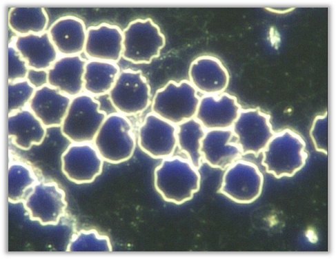
Poikilocytes are red blood cells that show variation in shape, rather than maintaining a consistent, round outline. In a live blood sample, this may include cells that appear elongated, irregular, angular, teardrop-shaped, or otherwise distorted compared with surrounding red blood cells.
In live blood analysis, it is common to observe an occasional irregularly shaped red blood cell within a field of view. This is generally considered a normal finding. Poikilocytosis becomes more relevant when multiple red blood cells within the same field, or across several fields, display noticeable shape variation rather than appearing as isolated, individual cells.
When shape variation is widespread, it suggests that red blood cell formation, maintenance, or survival within circulation may be less uniform than expected. The emphasis in live blood analysis is not on identifying a single cell type, but on recognising the overall pattern and proportion of irregular shapes present within the sample.
Pleomorphic Perspective
These forms develop as a result of consumption of erythrocyte material by the endobiont and indicate a high degree of endobiosis. Advanced poikilocytes (B above) are RBCs with sporoid symprotit inclusions. They appear as luminous spheres in darkfield and can occur in the RBC membrane, WBCs, or freely in plasma. When 80% of RBCs display this morphology, it is a dangerous sign. Sporoid symprotits in high numbers represent the upward development of the life cycle of Mucor racemosus Fresen and indicate pH imbalance and insufficient regulation.
Medical Perspective
Poikilocytosis is a general term used for abnormally shaped erythrocytes. A deviation from the normal discoid shape is due to chemical or physical alterations in the erythrocyte membrane or contents. Poikilocytosis includes all erythrocyte deformities (e.g., acanthocytes, codocytes, echinocytes, elliptocytes, spherocytes, stomatocytes, dacryocytes, keratocytes, microspherocytes, pyropoikilocytes, schistocytes, and semilunar bodies).
Relevance
Some degree of poikilocytosis will be present in all samples. This finding becomes more significant when observed in great numbers, exceeding 10% or more than 2–3 per viewing field. The damage progresses from RBCs with mildly irregular undulating edges (A above) to very irregular cells that appear to have black spots on them (B above). These severely damaged cells are often observed in high numbers in smokers and indicate a severe level of toxicity. This appearance can also be produced during the sampling procedure if the blood is exposed to alcohol that was not wiped off the finger, if the blood was overexposed to air (for longer than 10 seconds), or if the cover slip is pressurised. Always consider the whole sample and the patient — if in doubt, take another sample.
Implications
- Poikilocytes are formed as a result of lipid peroxidation (free radical damage) to the RBC membrane (A above). The more severely damaged RBC (B above) develops when haemoglobin within the RBC becomes damaged. These denatured haemoglobin aggregates then become attached to the inside of the RBC membrane.
- Considered a strong sign of toxicity and the need for detoxification.
- Deficiency of essential fatty acids & antioxidants.
- Dehydration. This picture is often observed in individuals who do not drink enough water and who’s only fluid intake constitute coffee, tea and soft drinks. Simply increasing water intake to optimum levels and restricting caffeine and alcohol intake will make a significant impact.
- Tobacco & environmental chemicals.
- Drugs/medication.
- Unbalanced diet, high in unhealthy fats, chemical additives (junk food).
- Carbonated beverages.
Some consider the “spotted” appearance of advanced poikilocytes to be due to Mycoplasmas on the RBC membrane. These are thought to be related to stress, low immunity, infections and possible rheumatoid arthritis. After having viewed a large number of specimens and studying the findings in their clinical context I agree with the researchers who include this finding under poikilocytosis as it is always present in advanced states of toxicity and mildly poikilocytic cells can be seen transforming into the advanced forms when studying the sample over the course of a few hours. Researchers who have arrived at this conclusion sometimes refer to the black spots as “Heinz Bodies”, which should not be confused with the Heinz Bodies observed in dry blood samples. The Heinz Bodies on RBC membranes occur when haemoglobin within the cell becomes damaged, precipitates and becomes attached the inner surface of the RBC membrane. In this manual, these severely damaged RBCs are referred to as “advanced poikilocytes”.
Associated Symptoms
- Fatigue
- A myriad of symptoms related to toxicity (headaches, malaise, skin conditions, etc.)
- Accelerated signs of aging
Interventions
Any combination of the following, depending on the rest of the case:
DETOX PROTOCOL:
Hepaton + Lymphlux + Nephrocil + HumiCaps + detox diet.
SUPPLEMENTS.
Omega-3 supplement (1000-2000 EPA daily).
Vitamin E: start with 400mg daily and gradually increase to 800mg daily.
Buffered vitamin C (2500mg) and/or other antioxidants such as: Super Oxide Dismutase, proanthocyanidins, N-Acetyl
Cysteine, selenium (200ug), beta carotene, zinc and glutathione.
Trace minerals: Bio-lonic Mineral Concentrate
Working with
As this finding indicates toxicity, the treatment focus is on detoxification, antioxidants, and adequate hydration. Most clients with significant poikilocytes do not drink enough water and consume excessive coffee/alcohol. Lymphatic toxins can also contribute (look for lymphatic patterns in dry blood samples, layers 1–4). Poor toxin elimination due to constipation, dehydration, and impaired liver function may also be observed. In such cases, treatment should address these factors. Investigate possible sources of toxicity such as household or pesticide exposure, even in ‘healthy’ raw foods.
General Guidelines
-
Moderate, or exclude, animal protein intake, aiming for balanced portions appropriate to body size and activity level
-
Avoid heavy or complex meal combinations; eat slowly, seated, and chew thoroughly to support digestive efficiency
-
Where relevant, consider individualised nutritional approaches that recognise biochemical uniqueness
-
Be mindful of potential food sensitivities or intolerances that may influence systemic balance
-
Support plasma fluidity by maintaining adequate daily water intake, adjusted for body weight and environment
-
Emphasise fibre-rich carbohydrates, leafy greens, sprouts, raw or lightly processed vegetables, and antioxidant-rich whole foods
-
Reduce reliance on highly processed foods, refined carbohydrates, and excessive saturated fats
-
Minimise or avoid smoking, alcohol, excess caffeine, refined sugar, and unnecessary chemical exposure where possible
Functional Systems Influenced
Hematological
Poikilocytes reflect variation in red blood cell shape within the bloodstream. Because red blood cells function most efficiently when they are uniform and flexible, noticeable shape variation highlights reduced consistency in red blood cell structure and behaviour.
Digestive & Nutrient Assimilation
Healthy red blood cell formation depends on the steady availability of nutrients required for cell development and membrane stability. When digestion or nutrient assimilation is less efficient, red blood cells may develop with greater variation in shape over time.
Metabolic
Red blood cell renewal and maintenance are metabolically driven processes. Reduced metabolic efficiency can influence how consistently red blood cells are formed and cleared, allowing structurally varied cells to persist in circulation.
Circulation & Hydration
Irregularly shaped red blood cells may move less smoothly through small blood vessels, particularly in areas where efficient microcirculation is essential. This can influence overall circulation quality, especially when fluid balance is suboptimal.
Commonly Associated Terrain Imbalances
Malabsorption / enzyme deficiency
When digestion or nutrient breakdown is less efficient, the body may not fully access the components required for consistent red blood cell formation. This can be reflected in increased variation in red blood cell shape.
Iron insufficiency
Iron plays a central role in red blood cell development and internal structure. Changes in iron availability or utilisation may be reflected in altered red blood cell morphology, without necessarily indicating a clinical deficiency.
B12 / Folate insufficiency
These nutrients support red blood cell maturation and structural consistency. Reduced availability over time may contribute to greater variability in red blood cell shape.
Mineral deficiency
Minerals act as cofactors in many cellular processes involved in red blood cell stability and renewal. Insufficiency may subtly influence cell shape consistency.
Protein intake / albumin low
Adequate protein availability supports plasma balance and red blood cell structure. Reduced availability may influence the consistency and resilience of red blood cell formation.
Supportive Focus & Awareness
-
Awareness of digestive efficiency and how well nutrients are being absorbed over time
-
Awareness of long-term nutrient availability that supports healthy red blood cell formation
-
Awareness of metabolic balance and its role in cellular renewal and consistency
-
Awareness of hydration and circulation quality, particularly at the microcirculatory level
-
Awareness of overall energy levels, stamina, and recovery capacity
Commonly Reported Experiences
Some individuals whose blood patterns include noticeable variation in red blood cell shape report feeling more easily fatigued or experiencing reduced stamina, particularly during periods of physical or mental demand. Others may notice slower recovery after exertion or a general sense of reduced resilience.
These experiences are non-specific and can be influenced by many factors, including lifestyle, stress, hydration, and nutritional status. Their presence does not confirm any condition and should always be considered in the broader context of individual circumstances and other observations.
Systems / Body Functions
Circulation & Hydration, Digestive & Nutrient Assimilation, Hematological, Metabolic
Imbalances
B12 / Folate insufficiency, Iron insufficiency, Malabsorption / enzyme deficiency, Mineral deficiency, Protein intake/albumin low
Poikilocytes are red blood cells that show variation in shape, rather than maintaining a consistent, round outline. In a live blood sample, this may include cells that appear elongated, irregular, angular, teardrop-shaped, or otherwise distorted compared with surrounding red blood cells.
In live blood analysis, it is common to observe an occasional irregularly shaped red blood cell within a field of view. This is generally considered a normal finding. Poikilocytosis becomes more relevant when multiple red blood cells within the same field, or across several fields, display noticeable shape variation rather than appearing as isolated, individual cells.
When shape variation is widespread, it suggests that red blood cell formation, maintenance, or survival within circulation may be less uniform than expected. The emphasis in live blood analysis is not on identifying a single cell type, but on recognising the overall pattern and proportion of irregular shapes present within the sample.
These forms develop as a result of consumption of erythrocyte material by the endobiont and indicate a high degree of endobiosis. Advanced poikilocytes (B above) are RBCs with sporoid symprotit inclusions. They appear as luminous spheres in darkfield and can occur in the RBC membrane, WBCs, or freely in plasma. When 80% of RBCs display this morphology, it is a dangerous sign. Sporoid symprotits in high numbers represent the upward development of the life cycle of Mucor racemosus Fresen and indicate pH imbalance and insufficient regulation.
 Disclaimer