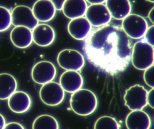
WBCs that are infected with pathogenic organisms. These are usually high valence symprotits that can be seen moving around actively in the WBC cytoplasm.
During the parasitic infestation of the WBC many different morphologies can emerge. Parasitised WBCs may have vacuoles in the cytoplasm, which will appear as dots of various sizes in the WBC that are white in brightfield and black in darkfield. WBCs can also develop dendritic growths extending out of the cell, which is an indication of advanced endobiosis. These dendritic growths are very thin and must be differentiated from the thicker, finger-like protrusions that the WBC uses to move (a favourable sign).
A common feature is that WBCs develop a translucent membrane, which is observed in viral conditions. When the translucent membrane contains many small nuclei, it is associated with a high degree of endobiosis.
When the parasitic burden becomes severe, the WBCs’ membranes rupture and their contents can be seen spilling out into the plasma (lysis). According to pleomorphism, leukaemia is the most extreme expression of WBC parasitism. Here the bone marrow, which is believed to be the workshop of the endobiont, has become infested with high valence microorganisms, resulting in the production of abnormal, leukemic WBCs.
Appearance
WBCs that are infected with pathogenic organisms. These are usually high valence symprotits that can be seen moving around actively in the WBC cytoplasm.
During the parasitic infestation of the WBC many different morphologies can emerge. Parasitised WBCs may have vacuoles in the cytoplasm, which will appear as dots of various sizes in the WBC that are white in brightfield and black in darkfield. WBCs can also develop dendritic growths extending out of the cell, which is an indication of advanced endobiosis. These dendritic growths are very thin and must be differentiated from the thicker, finger-like protrusions that the WBC uses to move (a favourable sign).
A common feature is that WBCs develop a translucent membrane, which is observed in viral conditions. When the translucent membrane contains many small nuclei, it is associated with a high degree of endobiosis.
When the parasitic burden becomes severe, the WBCs’ membranes rupture and their contents can be seen spilling out into the plasma (lysis). According to pleomorphism, leukaemia is the most extreme expression of WBC parasitism. Here the bone marrow, which is believed to be the workshop of the endobiont, has become infested with high valence microorganisms, resulting in the production of abnormal, leukemic WBCs.
Relevance
Parasitised WBCs is not a normal finding and is considered significant when observed during analysis. These anomalies may not be obvious at the beginning of the analysis and may only emerge after some time.
Implications
Parasitized WBCs indicate a high degree of parasitism by the pathogenic forms of the endobiont due to an unbalanced terrain. Look at the different expressions of the higher growth forms of the endobiont in the WBCs, RBCs and plasma to determine the degree of endobiontic burden. Remember that the progression of the endobiont into its higher, pathogenic growth forms occur because of an unbalanced terrain. The higher growth forms require a progressively lower (acidic) pH and unbalanced redox potential (hydrogen-ion concentration).
Associated Symptoms
- Fatigue
- Skin and mucous membrane yeast and fungal infections.
- Susceptible to colds and flu.
 Disclaimer