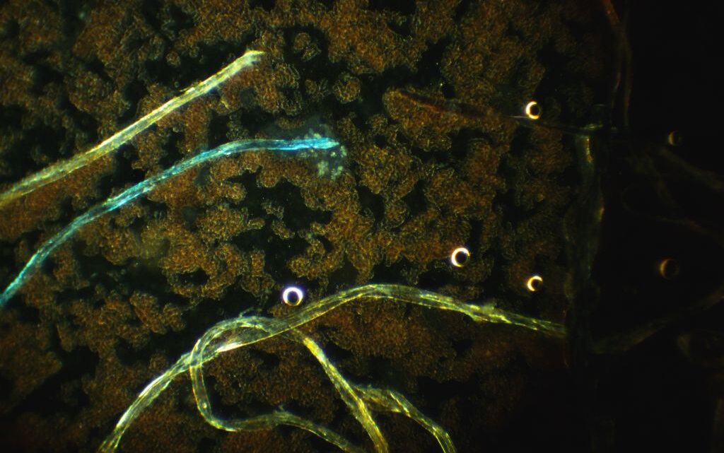
This darkfield micrograph shows elongated, thread-like structures likely identified as nematode larvae, likely from a species such as Strongyloides or Ancylostoma, against a background of heavily aggregated red blood cells (RBCs). The larvae are bright and sinuous, with a smooth, continuous form, ranging from 200 to 600 micrometres in length depending on the species.
The RBC aggregation, appearing as dense clusters or rouleaux formation, may be influenced by the parasitic infection, potentially due to inflammation or altered plasma proteins caused by the larvae. The circular, ring-like objects scattered throughout are consistent with additional RBCs or air bubbles, typical in such preparations. The presence of larvae suggests a parasitic infection, which would require further clinical correlation for confirmation.
The life cycle of *Strongyloides stercoralis*, a common parasitic nematode, is complex and includes both free-living and parasitic stages. Here are the key details:
Infective Stage: The cycle begins with the free-living infective third-stage filariform larvae (L3), which penetrate human skin, typically through contact with contaminated soil. This stage is non-feeding and adapted for host entry.
Migration to Lungs: Once inside, larvae enter the bloodstream and are carried to the lungs. They penetrate the alveoli, ascend the bronchial tree, and are coughed up and swallowed, reaching the small intestine.
Maturation in Intestine: In the small intestine, the larvae mature into adult female worms (males are rare or absent in humans). These parthenogenetic females (1-2 mm long) embed in the intestinal mucosa, particularly the duodenum and jejunum, and produce eggs via self-fertilisation.
Egg Hatching: Eggs hatch within the intestine into rhabditiform first-stage larvae (L1), which are excreted in faeces. This stage feeds on intestinal contents or external organic matter if in soil.
Free-Living Cycle (Optional): In favourable conditions (e.g., warm, moist soil), excreted rhabditiform larvae can develop into free-living adults outside the host. These adults (both male and female) mate, and the females lay eggs that hatch into rhabditiform larvae, which then moult into infective filariform larvae (L3), restarting the cycle.
Autoinfection: Inside the host, some rhabditiform larvae can moult into filariform larvae in the intestines without being excreted. These penetrate the intestinal wall or perianal skin, re-entering the circulation to repeat the migration (lung-intestine cycle), enabling chronic infection.
Duration: The prepatent period (time from infection to egg production) is about 2-4 weeks. Without autoinfection, the parasitic phase lasts 2-5 years, but autoinfection can lead to decades-long infections.
Environmental Factors: The free-living cycle thrives in tropical and subtropical climates with adequate moisture and temperature (20-30°C), while autoinfection is more common in immunocompromised individuals, potentially causing hyperinfection syndrome.
This dual life cycle allows *Strongyloides* to persist in both human hosts and the environment, making it a persistent health concern, especially in endemic areas.
 Disclaimer