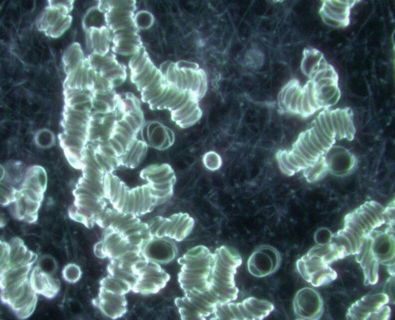

In this image, we can see that the red blood cells are not moving freely and independently, but instead are gathering into small chains and clumps — a process known as aggregation. While this is significant in itself, the most striking feature here is not the cells but the delicate web that surrounds them. Those fine, hair-like strands stretching between and around the clusters are fibrin.
Fibrin is a fibrous protein produced when fibrinogen, a soluble protein in plasma, is converted as part of the body’s natural clotting response. Under normal conditions, fibrin plays a vital role in repairing wounds by creating a mesh that stops bleeding and helps tissue heal. However, when we observe fibrin in circulating blood — away from an injury site — it can point toward other processes at work. Its presence may indicate underlying inflammation, an environment where the blood has become sticky or sluggish, or a tendency toward excessive clotting activity.
Viewed under darkfield microscopy, fibrin takes on the appearance of a ghostly cobweb or network of fine threads. It creates a background texture that contrasts sharply with the otherwise dark field, linking the red cells together and reducing their ability to flow smoothly. This can restrict oxygen delivery and circulation, highlighting how even subtle changes at the microscopic level can influence the body’s overall vitality and resilience.
 Disclaimer